Arteries Diagram / Artery And Vein Diagram For Powerpoint Pslides
Arteries Diagram / Artery And Vein Diagram For Powerpoint Pslides. Blockage of the posterior inferior cerebellar artery can result in a type of stroke called lateral medullary syndrome. Arteries and veins are two of the body's main type of blood vessels. When this happens, less blood flows to your legs. The coronary arteries wrap around the outside of the heart. We think this is the.
ads/bitcoin1.txt
Systemic arteries deliver blood to the rest of the body. Within the cranial vault, the terminal branches of these arteries form an anastomotic circle, called the circle of willis.from this circle, branches arise which supply the majority of the. Main branches from the aorta include the brachiocephalic artery, left carotid artery, and the left subclavian artery. When this happens, less blood flows to your legs. The right common carotid artery arises from a bifurcation of the brachiocephalic trunk (the right subclavian artery is the other branch).
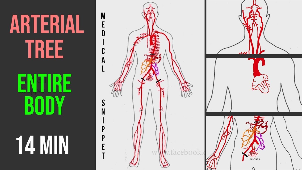
Arteries Arterial Tree Of The Entire Human Body Anatomy Explained In 14 Minutes Youtube from i.ytimg.com Coronary circulation is the circulation of blood in the blood vessels that supply the heart muscle (myocardium). Original vintage human anatomy victorian bookplate print 1890s medical diagram veins arteries blood circulatory system of the human body thepapermuseum. Diagram of the human circulatory system (infographic). If your leg arteries are badly blocked, you may develop foot pain while resting or a sore that won't heal. When this happens, less blood flows to your legs. Circle of willis is indeed a hot neuroanatomy topic! Blood carried by arteries is usually highly oxygenated, having just left the lungs on its way to the body's tissues. There are two paired arteries which are responsible for the blood supply to the brain;
The right common carotid artery arises from a bifurcation of the brachiocephalic trunk (the right subclavian artery is the other branch).
ads/bitcoin2.txt
It originates from the heart and branches out into smaller arteries which supply blood to the head region (brachiocephalic artery), the heart itself (coronary arteries), and the lower regions of the body. Because the rest of the body, and most especially the brain, needs a steady supply of oxygenated blood that is free of all but the slightest. Most arteries carry oxygenated blood; Arteries are the blood vessels that carry blood away from the heart, where it branches into even smaller vessels. An artery (plural arteries) (from greek ἀρτηρία (artēria) 'windpipe, artery') is a blood vessel that takes blood away from the heart to one or more parts of the body (tissues, lungs, brain etc.). Learn the differences between an artery and a vein. We shall start at the origin of the carotid arteries. Coronary circulation is the circulation of blood in the blood vessels that supply the heart muscle (myocardium). When this happens, less blood flows to your legs. The arteries in your legs and feet can get blocked, just like the arteries in your heart. The system is responsible for the flow of blood, nutrients. 5 out of 5 stars (293) 293 reviews $ 24.27. When the coronary arteries narrow to the point that blood flow to the heart muscle is limited (coronary artery disease), collateral vessels may enlarge and become active.
When this happens, less blood flows to your legs. It is one of the three main arteries that supply blood to the cerebellum , a part of the brain. These vessels are channels that distribute blood to the body. Ascending aorta, aortic arch, thoracic aorta, and abdominal aorta. This allows blood to flow around the blocked artery to another artery nearby or to the same artery past the blockage, protecting the heart tissue from injury.
Fully Labeled Arteries Diagram Jpg Superficial Temp Oral Artery Posteria Auricular Artery External Carotid Artery Internal Carotid Artery Common Course Hero from www.coursehero.com It originates from the heart and branches out into smaller arteries which supply blood to the head region (brachiocephalic artery), the heart itself (coronary arteries), and the lower regions of the body. The aorta is the main systemic artery and the largest artery of the body. Resistance (r) the force opposing blood flow. Arteries carry blood away from the heart in two distinct pathways: The tunica medica, which is the very muscular middle layer in arteries, is thinner and less muscular in veins. We think this is the. We shall start at the origin of the carotid arteries. It is a central communication that unites the internal carotid and vertebrobasilar systems.
Arteries are blood vessels that carry blood away from the heart.
ads/bitcoin2.txt
When the coronary arteries narrow to the point that blood flow to the heart muscle is limited (coronary artery disease), collateral vessels may enlarge and become active. Most arteries carry oxygenated blood; We shall start at the origin of the carotid arteries. Lungs (pulmonary), and arteries, veins, coronary and portal vessels (systemic). Arteries are the blood vessels that carry blood away from the heart, where it branches into even smaller vessels. Anatomynote.com found human body artery diagram in detail from plenty of anatomical pictures on the internet. This allows blood to flow around the blocked artery to another artery nearby or to the same artery past the blockage, protecting the heart tissue from injury. An artery (plural arteries) (from greek ἀρτηρία (artēria) 'windpipe, artery') is a blood vessel that takes blood away from the heart to one or more parts of the body (tissues, lungs, brain etc.). Circle of willis is indeed a hot neuroanatomy topic! The subclavian arteries turn unto the brachial arteries as they pass through the upper arm which feed the radial and ulnar arteries. The veins also lack the elastic internal lamina that lies. Coronary arteries supply blood to the heart muscle. The system is responsible for the flow of blood, nutrients.
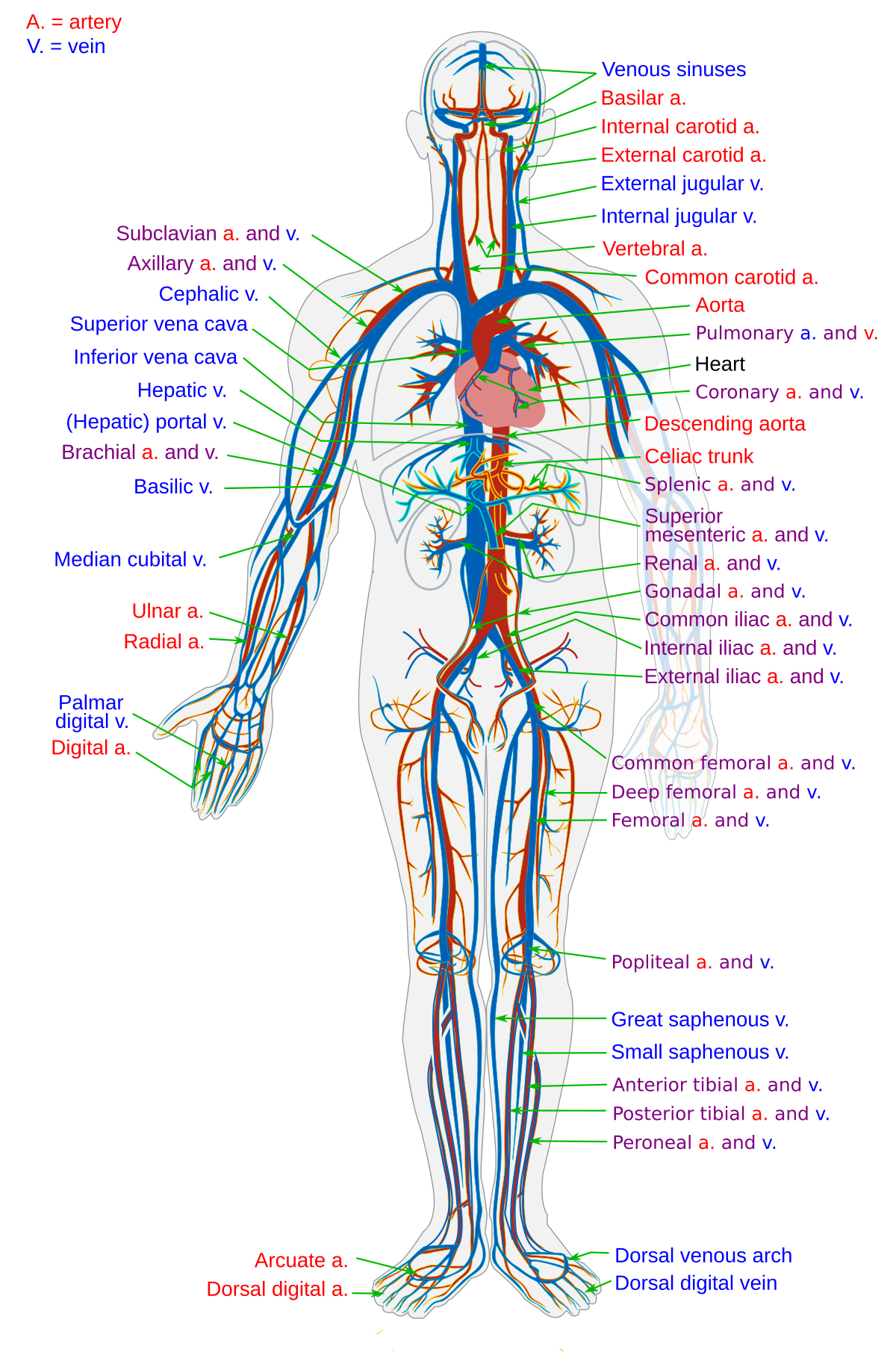
There is a point at which the anterior and posterior arterial circuits of the brain unite or anastomose. Learn the differences between an artery and a vein. Anatomynote.com found human body artery diagram in detail from plenty of anatomical pictures on the internet. Anatomynote.com found blood circulation principal veins and arteries diagram from plenty of anatomical pictures on the internet. These vessels are channels that distribute blood to the body.
Blood Vessel Wikipedia from upload.wikimedia.org These arteries arise in the neck, and ascend to the cranium. Arteries carry blood away from the heart in two distinct pathways: Systemic arteries deliver blood to the rest of the body. Cholesterol plaques can be the cause of heart disease. Most arteries carry oxygenated blood; Plaques begin in artery walls and grow over years. This allows blood to flow around the blocked artery to another artery nearby or to the same artery past the blockage, protecting the heart tissue from injury. Circle of willis is indeed a hot neuroanatomy topic!
After receiving blood directly from the left ventricle of the heart, the.
ads/bitcoin2.txt
A condition which arises spontaneously or as the result of trauma, where the walls of the artery are split, leading to internal bleeding and disruption of blood flow. Coronary circulation is the circulation of blood in the blood vessels that supply the heart muscle (myocardium). Within the cranial vault, the terminal branches of these arteries form an anastomotic circle, called the circle of willis.from this circle, branches arise which supply the majority of the. This allows blood to flow around the blocked artery to another artery nearby or to the same artery past the blockage, protecting the heart tissue from injury. For more anatomy content please follow us and visit our website: Blood is pumped from the heart in the arteries. When the coronary arteries narrow to the point that blood flow to the heart muscle is limited (coronary artery disease), collateral vessels may enlarge and become active. The right common carotid artery arises from a bifurcation of the brachiocephalic trunk (the right subclavian artery is the other branch). Finally, the smallest arteries, called arterioles are further branched into small capillaries, where the exchange of all the nutrients, gases and other waste molecules are carried out. The capillaries connect the two types of blood. This area is known as the circle of willis. Coronary arteries supply blood to the heart muscle. The vertebral arteries, and the internal carotid arteries.
ads/bitcoin3.txt
ads/bitcoin4.txt
ads/bitcoin5.txt
0 Response to "Arteries Diagram / Artery And Vein Diagram For Powerpoint Pslides"


0 Response to "Arteries Diagram / Artery And Vein Diagram For Powerpoint Pslides"
Post a Comment